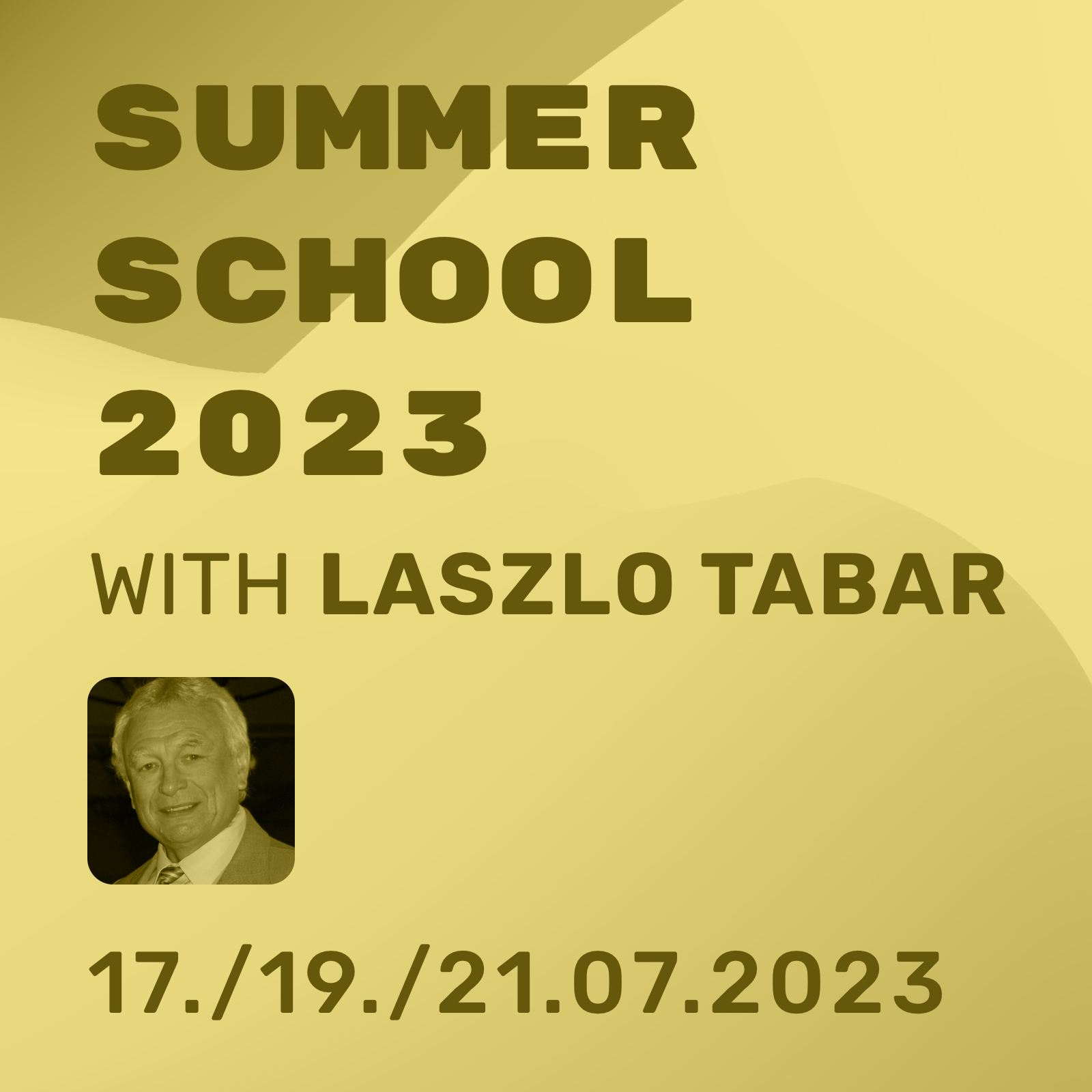
Summer School 2023

Summer School Breast Imaging
with Dr. László Tabár

A webinar-based, interactive, unique learning experience

17/19/21 July 2023, Online
Each time 18:00 to 21:00

This product is currently out of stock and unavailable.

SKU: N/A Categories: , ,

Description

Trailer

Want to learn more about our Summer School?

• Watch here the full trailer of László Tabár! Here you will:
• Learn the concept of the course
• Understand the objectives of each session
• Grasp the passion of Doctor Tabar´s which drives his unparalleled breast imaging teaching.

Please also have a look on the flyer for this years Summer School 2023!

Trailer about the Summer School 2023
László Tabár

Dear colleagues,

With 750+ registrants from 75+ countries around the globe our spring “Mammography Symposium” with László Tabár was a huge success.

We are more than happy to announce that our friend László Tabár agreed to join us again!

Doctor Tabár will hold a Summer School specifically designed to improve your skills in reading and interpreting breast images.

This interactive webinar-based course, presented by THE authority in the field of breast imaging, will provide you a unique learning experience.

In our Summer school you will receive extensive knowledge about breast imaging, differential diagnosis of breast diseases, and their implications for patient management

You will find all necessary information and background on this page. Please check also our flyer and the trailer video by Doctor Tabár.

See you soon!

Yours,
Pascal Baltzer and Matthias Dietzel

Directors, School of Radiology, organizers

Outline

1

Mix of impulse lecture and interactive case reading session: Lectures on each major subject will be mixed with interactive sessions. These consist of questions relevant to the subject and a mixture of normal and early cancer cases presented as they appear on a viewing station.
2

Using interactive online polls, your personal learning progress will be evident. This course design gives immediate feedback demonstrating the effectiveness of our teaching methods.
3

During the course you will progressively improve your skills in reading and interpreting breast images.
4

You will learn the full spectrum of normal breast images, with all important findings explained with the help of 3-dimensional histology images.
5

Our course aims at helping you to reduce unnecessary call-backs and increase your confidence in reading mammograms.
6

You will receive immediate feedback and histologic confirmation using large format histology.
7

The multimodality approach will be emphasized throughout the course.
8

Special emphasis will be placed on finding early phase breast cancers.

Concept

1

Three evening sessions, 2.5 hours each
2

Video will be shared with you after the course
3

9 CME credits (applied, 3 per session)
4

Engaging and motivating learning atmosphere
5

Latest e-learning technology including HD-streaming and online live polls.
6

Real time interaction with your tutors and peers
7

The multimodality approach will be emphasized throughout the course.
8

Special emphasis will be placed on finding early phase breast cancers.

Objectives

This course provides extensive knowledge about diagnostic breastimaging. Key objectives are to …

1

Improve skills in reading and interpreting breast images
2

Identify normal examinations
3

Understand typical patterns of malignant findings
4

Practice efficient and effective reporting techniques
5

Use multimodal breast imaging in a rational way
6

Apply differential diagnosis of breast diseases
7

Approach Implications for patient management

Target audiences

1

Our course aims at all professionals involved in breast cancer care such as:
Radiologists, Surgeons, Pathologists, Gynecologists, and Radiology Technologists
2

Our course is designed for all levels of expertise in breast imaging:
Ranging from Level 1 (beginners), 2 (Intermediate), 3 (advanced), and 4 (experts).

What our par­tic­i­pants say

Programme Day 1

Monday, 17.07.2023

Topic
Content & Speaker

18:30

Welcome

Didactic lectures

Tabár, moderators: Baltzer, Dietzel

18:40

A new era in the diagnosis and treatment of breast cancer. A short history.

  • Correlating 3-dimensional, subgross anatomy with mammography of the normal breast results in increased confidence in reading a mammogram and finding small abnormalities.
  • Special training in large format thin and thick section (3D) histopathologic correlation enables the radiologist to account for every linear and nodular density on the mammogram.

Finding invasive breast cancer when it is still small

  • Malignant stellate and circular/oval-shaped lesions originating from the TDLUs (AAB):
  • Clinical presentation, histology, mammographic – MRI – ultrasound appearance and outcome.

A systematic method for viewing mammograms

  • Areas on the mammogram where most breast cancers will be found.
  • Viewing dense breasts
  • Viewing relatively easy to-read breasts

The multimodality approach

The role of hand-held ultrasound / 3D automated ultrasound / MRI in the detection and workup of the findings.

Algorithm for classifying breast diseases according to their site of origin

19:45

Break

Interactive session

Tabár, moderators: Baltzer, Dietzel

20:00

Cases reading

  • Each participant will assess a mixture of normal and early cancer cases using an interactive online voting system
  • Results will appear instantly for discussion and evaluation.
  • All abnormal cases are fully worked up
  • Complete imaging workup will be presented in detail, including ultrasound, MRI and large section histopathology.

20:50

Wrap up

21:00

End of the event

Programme Day 2

Wedesday, 19.07.2023

Topic
Content & Speaker

18:30

Welcome

Didactic lectures: Calcification on the mammogram

Tabár, moderators: Baltzer, Dietzel

18:40

Algorithm for classifying breast diseases according to their site of origin

  • Breast diseases originating in the major ducts
  • Benign type calcifications originating in the major ducts
  • Secretory disease type calcifications
  • Malignant type calcifications originating in the major ducts

19:45

Break

Interactive session

Tabár, moderators: Baltzer, Dietzel

20:00

Cases reading

  • Each participant will assess cases using an interactive online voting system
  • Results will appear instantly for discussion and evaluation.
  • All abnormal cases are fully worked up
  • Complete imaging workup will be presented

20:50

Wrap up

21:00

End of the event

Programme Day 3

Friday, 21.07.2023

Topic
Content & Speaker

18:30

Welcome

Didactic lectures

Tabár, moderators: Baltzer, Dietzel

18:40

Algorithm for classifying breast diseases according to their site of origin

  • Benign breast diseases originating in the TDLU and associated with calcifications on the mammogram:
  • Fibrocystic change
  • Fibroadenoma
  • Different types of adenosis
  • Understanding pathophysiology leading to calcified and non-calcified hyperplastic breast changes.
  • The morphologic analysis of calcifications representing a less aggressive carcinoma: Grade 1 / well differentiated CIS
  • Mammographic / histopathologic correlation of pleomorphic calcifications representing Grade 2 CIS within the TDLU

19:45

Break

Interactive session

Tabár, moderators: Baltzer, Dietzel

20:00

Cases reading

  • Each participant will assess cases using an interactive online voting system
  • Results will appear instantly for discussion and evaluation.
  • All abnormal cases are fully worked up
  • Complete imaging workup will be presented

20:50

Wrap up

21:00

End of the event

Literature

Tabár L, Dean PB, Tucker FL, et al (2022) A new approach to breast cancer terminology based on the anatomic site of tumour origin: The importance of radiologic imaging biomarkers. European Journal of Radiology 149: https://doi.org/10.1016/j.ejrad.2022.110189

Tabár L, Dean PB, Tucker FL, et al (2022) Breast cancers originating from the terminal ductal lobular units: In situ and invasive acinar adenocarcinoma of the breast, AAB. European Journal of Radiology 152: https://doi.org/10.1016/j.ejrad.2022.110323

Tabár L, Dean PB, Tucker FL, et al (2022) Breast cancers originating from the major lactiferous ducts and the process of neoductgenesis: Ductal Adenocarcinoma of the Breast, DAB. European Journal of Radiology 153: https://doi.org/10.1016/j.ejrad.2022.110363

Tabár L, Dean PB, Tucker FL, et al (2022) Imaging biomarkers of breast cancers originating from the major lactiferous ducts: Ductal adenocarcinoma of the breast, DAB. European Journal of Radiology 154: https://doi.org/10.1016/j.ejrad.2022.110394

Tabár L, Dean PB, Tucker FL, Vörös A (2023) Can we improve breast cancer management using an image-guided histopathology workup supported by larger histopathology sections? European Journal of Radiology 161: https://doi.org/10.1016/j.ejrad.2023.110750

Tabár L, Dean PB, Tucker FL, et al (2023) The challenging imaging and histopathologic features of diffusely infiltrating breast cancer. European Journal of Radiology 161: https://doi.org/10.1016/j.ejrad.2023.110754

Sponsorship / Conflicts of Interest

Our mission is: Continued medical education by radiologists for radiologists. We do not receive financial contributions from third parties (sponsoring, industry, etc.). This course is financed exclusively by the fees of the participants.

Venue: Online (Zoom)

Access links and technical instructions will be emailed to participants 3 days prior to the start of the event.

Pricing

Ticket
Price

Summer School 2023

3-days-ticket

299,00 € incl. VAT

2-days-ticket *

219,00 € incl. VAT

1-day-ticket *

139,00 € incl. VAT

Discounts

Resident

-30%

Bundle **

-20%

Video only ***
Only upon prior request:
Send us an email

-20%

* We strongly recommend to join the complete Summer School, as it is designed as a coherent course.
In case you are not able to join the complete course, you may book one or two sessions separately. Note that every attendee will also get access to the videos. Videos are courtesy by Doctor Tabar and will be available on his e-leanring platform. In case you still are not able to attend the full course: The following options are available:
1-day ticket: Each day may be booked separately.
2-day ticket: The following combos are approved by Doctor Tabar: Day 1+2, Day 2+3


** Applicable, if you book at least two different courses at the School of Radiology at the same time.


*** Interaction is key in this course. It is strongly recommended to join the summer school live. However, in case you cannot attend the course live, we provide a limited number of video-only tickets. These tickets are available on personal request only. If you are interested, please write to our office: office@school-radiology.com

FAQ

What emails will I receive and when ?

You will receive all necessary documents from us by email.
The following information will be e-mailed to you:

  • Immediately after your booking: booking confirmation + invoice + payment information (if you have chosen payment by invoice).
  • 3 days before your course: Detailed information + access key to your course.
  • Immediately after your course: Confirmation of participation.

Tip:
Please check the e-mail address that you provided for typos, and regularly check your inbox and junk mail folders.

I did not receive any e-mail from you!

Our emails are occasionally blocked by firewalls and junk mail filters. Unfortunately, we have no control over this. 
If you did not receive our e-mails, please reach out to us via e-mail, phone, instant messenger, or SMS:

+49 157 58741925
office@school-of-radiology.com

We will gladly assist you and resend the information!

Tip:
Check your junk mail folder.
Add our address to your e-mail whitelist.
Check the e-mail you provided for typos.

How do I book a course?

All course bookings are done directly on our webshop.
Select your course(s) at https://school-of-radiology.com/courses/
Put the course(s) in your shopping basket.
Follow the instructions provided.

Tip:
If you have any problems, please feel free to contact us directly (see: “How can I contact you directly?”).

Do you have discounts?

Good news: You can get a wide range of discounts!
All discounts are applied automatically.

  • Bundle discount:
    You can receive a 20% bundle discount when booking a minimum of two courses at the same time.
  • Resident’s discount:
    As a resident in training, you receive a 30% discount.
  • Combination:
    All discounts can be combined.

Tip:
Many of our courses are sequential (basic + advanced courses).
You can book them together to receive your discount!
Even experts will benefit from joining our basic courses.

How can I pay?

You can pay using a credit card, Paypal, or SEPA bank transfer. The amount will be debited automatically. We also accept manual payment by invoice.

Tip:
Our bank account information is not routinely included in the invoice.
Please copy the bank account details displayed at the end of the checkout process.

When will I receive my access code?

We will e-mail your access code to you 3 days before your course.

When will I receive my participation confirmation?

We will e-mail your participation confirmation immediately after your course.
In this file you will find:

  • Your personal participation confirmation
  • The number and confirmation of the CME credits you earned
  • Confirmation of the number and types of cases you assessed during the course (this is optional; see also: “Will I receive a certificate on my case assessments?”)

Will I receive a certificate for my case assessments?

In our full-day courses, you will independently assess real-life cases. You will use our online tool and apply structured reporting for this assessment. Each case is quality-controlled, and we will discuss your assessment.

In our full-day courses we are happy to certify the number of cases that you independently reported (note: this service is optional).

In this case you will find the certificate in your participation confirmation on the last page.

Where can I find your bank details?

We display our bank account at the end of the checkout process. Note that our bank details are not included in the invoice.

Tip:
If you have any questions, please contact us directly (FAQ “How can I contact you directly?”).

How do I receive my CME credits?

We will e-mail your CME certificate directly after the course (see: “When will I receive my confirmation of participation?”). All the necessary information about the type and amount of your CME credits will be included.

Tip:
Details on the CME credits are outlined on each course site.

What technical equipment do you recommend?

Our courses are held online via Zoom.
All you need is an internet-compatible device of your choice and a stable internet connection.
The following tips can maximize your learning experience:

  • Install Zoom on your computer.
  • Log in with your personal Zoom account.
  • Familiarize yourself with basic functionalities, e.g., turning video and audio on/off and writing chat messages.
  • Please do not use Zoom via your browser.
  • Tip: Install the Zoom app on a second device (smartphone, tablet, etc.). This will make answering interactive polls more convenient. It will also serve as a backup.
  • Choose a high-quality monitor. If possible, use your diagnostic workstation.
  • A fast and stable internet connection (ideally LAN, not W-LAN), appropriate lighting, a quality microphone, and a quality webcam are important elements for success in the course.

How can I contact you directly?

Our company does not have a hotline. We answer your questions personally.
You may also talk directly to your tutors for specific questions related to the course content.
So please contact us at any time by e-mail, phone, instant messenger, SMS, etc.:

+49 157 58741925
office@school-of-radiology.com

CME Accreditation

In our one day courses you will achieve optimal learning success and maximum CME points in the shortest possible time!

9

CME points (category A, applied for at the BLÄK); 3 credits per sessions

You want more?

Sure! If you book several courses at the same time, you receive a 20% discount on the total amount.

20

% discount

Resident in training?

We would like to make our contribution! We support your training with a 30% discount on all courses!

30

% discount

Quality assurance