
Description
Trailer
Want to learn more about our Summer School?
• Watch here the full trailer of László Tabár! Here you will:
• Learn the concept of the course
• Understand the objectives of each session
• Grasp the passion of Doctor Tabar´s which drives his unparalleled breast imaging teaching.
Please also have a look on the flyer for this years Summer School 2023!
Trailer about the Summer School 2023
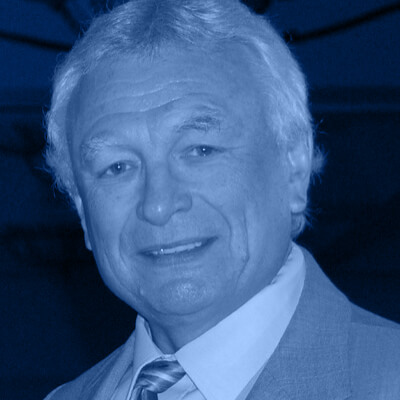
László Tabár

Dear colleagues,
With 65+ live attendees from 25+ countries around the globe our “Summer School with László Tabár“ was a huge success.
You missed the live course?
We are more than happy to announce that our friend László Tabár agreed to share the video edition of his course!
This 5.5h video course is specifically designed to improve your skills in reading and interpreting breast images.
In addition to the core program László Tabár has included exclusive bonus material in the video edition: Here you will learn from his additional great teaching cases and watch exclusive lectures.
This course by THE authority in the field of breast imaging, will provide you a unique learning experience.
Just like in the live version you will receive extensive knowledge about breast imaging, differential diagnosis of breast diseases, and their implications for patient management.
You will find all necessary information and background on this page. Please check also our flyer and the trailer video by Doctor Tabár.
In case of any questions, reach out to us anytime. We will be happy help!
See you soon
Yours,
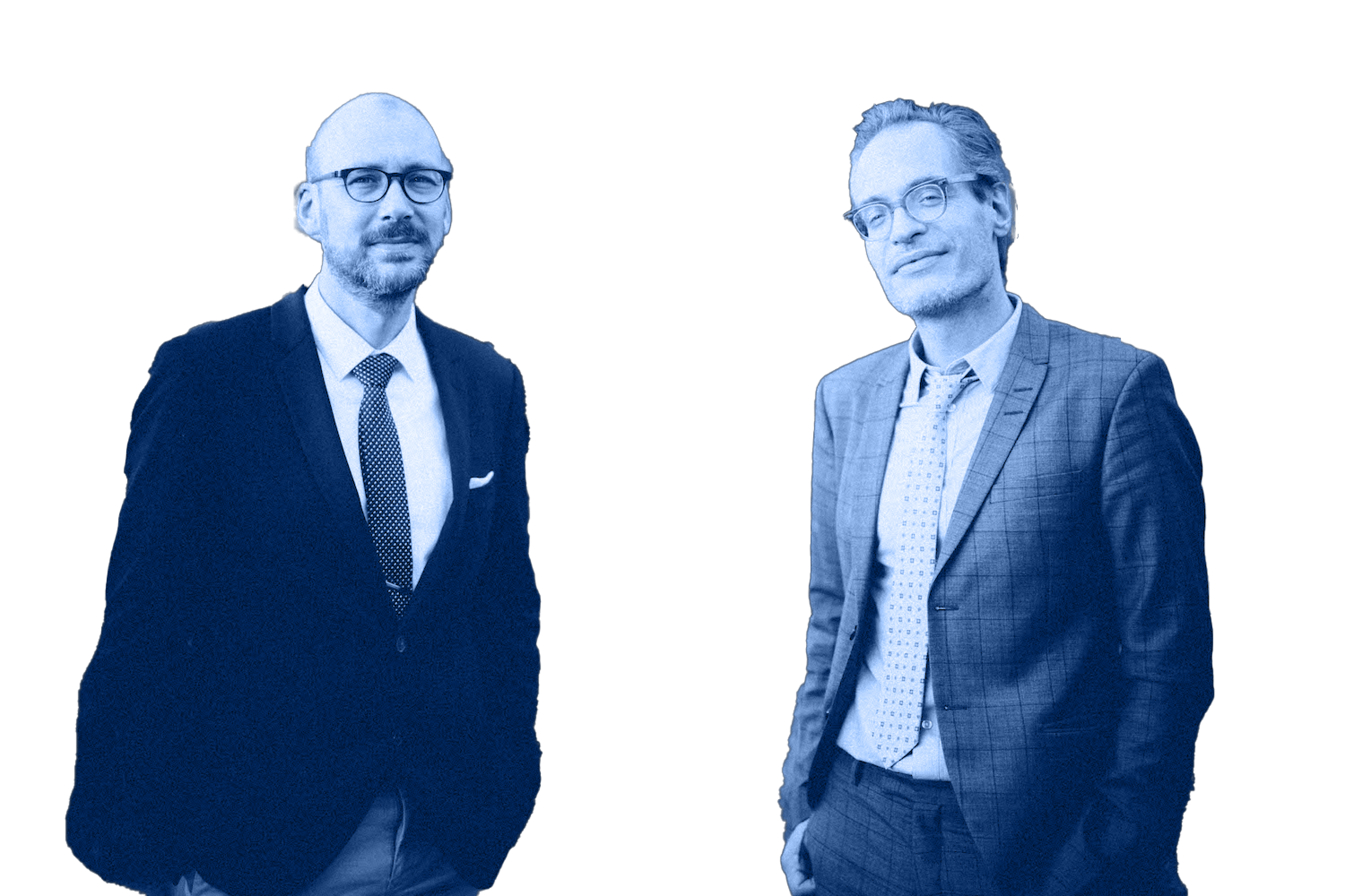
Pascal Baltzer and Matthias Dietzel
Directors, School of Radiology, organizers
Outline
Concept
Objectives
This course provides extensive knowledge about diagnostic breastimaging. Key objectives are to …
Target audiences
What our participants say
„
I participated in this years course to deepen my understanding of mammography.
I was deeply impressed by the insights, beauty and significance of the course. I wholeheartedly recommend this summer school event to everyone seeking deeper knowledge and understanding in the field of mammography.
László Tabar’s depth of knowledge and his exceptional ability to share that made a huge impression on me and I found it to be very insipiring.
All that leads me to say, that this has been – by far – the best online course and lecture I have ever experienced in my life. Thank you.
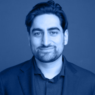
Muhammad Taha Hagar, MD
Radiology resident. University hospital Freiburg, Germany
„
Thank you so much for all your efforts making it possible to attend your most up to date courses with the best speakers. So that already being an experienced breast imager, I can still learn new things. Knowledge that leads to better outcomes and improved diagnosis for my patients. Everything is designed at the SOR to help us learn new skills at our own pace. Courses are suited to all skill levels, with so many tips and tricks to work. Although there is still a disparity between health systems around the world, you connect us all. Even, if we don’t have the latest equipment to our hand. So, everybody can learn! Every course is an enlightening and fun experience: Easy and simple to understand. And most: Every question is being answered. It is priceless to read a personal feedback only for me, with all the explanations what I asked and even more! Thank you, for the special opportunities the SOR has given to me.
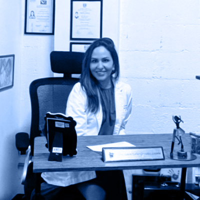
Laura Esther Teté Gonzalez Lozada, MD
Breast radiologist, private practice
Day 1
Topic
A new era in the diagnosis and treatment of breast cancer. A short history.
- Correlating 3-dimensional, subgross anatomy with mammography of the normal breast results in increased confidence in reading a mammogram and finding small abnormalities.
- Special training in large format thin and thick section (3D) histopathologic correlation enables the radiologist to account for every linear and nodular density on the mammogram.
Finding invasive breast cancer when it is still small
- Malignant stellate and circular/oval-shaped lesions originating from the TDLUs (AAB):
- Clinical presentation, histology, mammographic – MRI – ultrasound appearance and outcome.
A systematic method for viewing mammograms
- Areas on the mammogram where most breast cancers will be found.
- Viewing dense breasts
- Viewing relatively easy to-read breasts
The multimodality approach
The role of hand-held ultrasound / 3D automated ultrasound / MRI in the detection and workup of the findings.
Algorithm for classifying breast diseases according to their site of origin
Cases reading
- Each participant will assess a mixture of normal and early cancer cases using an interactive online voting system
- Results will appear instantly for discussion and evaluation.
- All abnormal cases are fully worked up
- Complete imaging workup will be presented in detail, including ultrasound, MRI and large section histopathology.
Day 2
Topic
Algorithm for classifying breast diseases according to their site of origin
- Breast diseases originating in the major ducts
- Benign type calcifications originating in the major ducts
- Secretory disease type calcifications
- Malignant type calcifications originating in the major ducts
Cases reading
- Each participant will assess cases using an interactive online voting system
- Results will appear instantly for discussion and evaluation.
- All abnormal cases are fully worked up
- Complete imaging workup will be presented
Day 3
Topic
Algorithm for classifying breast diseases according to their site of origin
- Benign breast diseases originating in the TDLU and associated with calcifications on the mammogram:
- Fibrocystic change
- Fibroadenoma
- Different types of adenosis
- Understanding pathophysiology leading to calcified and non-calcified hyperplastic breast changes.
- The morphologic analysis of calcifications representing a less aggressive carcinoma: Grade 1 / well differentiated CIS
- Mammographic / histopathologic correlation of pleomorphic calcifications representing Grade 2 CIS within the TDLU
Cases reading
- Each participant will assess cases using an interactive online voting system
- Results will appear instantly for discussion and evaluation.
- All abnormal cases are fully worked up
- Complete imaging workup will be presented
Literature
Tabár L, Dean PB, Tucker FL, et al (2022) A new approach to breast cancer terminology based on the anatomic site of tumour origin: The importance of radiologic imaging biomarkers. European Journal of Radiology 149: https://doi.org/10.1016/j.ejrad.2022.110189
Tabár L, Dean PB, Tucker FL, et al (2022) Breast cancers originating from the terminal ductal lobular units: In situ and invasive acinar adenocarcinoma of the breast, AAB. European Journal of Radiology 152: https://doi.org/10.1016/j.ejrad.2022.110323
Tabár L, Dean PB, Tucker FL, et al (2022) Breast cancers originating from the major lactiferous ducts and the process of neoductgenesis: Ductal Adenocarcinoma of the Breast, DAB. European Journal of Radiology 153: https://doi.org/10.1016/j.ejrad.2022.110363
Tabár L, Dean PB, Tucker FL, et al (2022) Imaging biomarkers of breast cancers originating from the major lactiferous ducts: Ductal adenocarcinoma of the breast, DAB. European Journal of Radiology 154: https://doi.org/10.1016/j.ejrad.2022.110394
Tabár L, Dean PB, Tucker FL, Vörös A (2023) Can we improve breast cancer management using an image-guided histopathology workup supported by larger histopathology sections? European Journal of Radiology 161: https://doi.org/10.1016/j.ejrad.2023.110750
Tabár L, Dean PB, Tucker FL, et al (2023) The challenging imaging and histopathologic features of diffusely infiltrating breast cancer. European Journal of Radiology 161: https://doi.org/10.1016/j.ejrad.2023.110754
Sponsorship / Conflicts of Interest
Our mission is: Continued medical education by radiologists for radiologists. We do not receive financial contributions from third parties (sponsoring, industry, etc.). This course is financed exclusively by the fees of the participants.
Pricing
Option
Summer School 2024
Video Edition
149,50 € incl. VAT
Summer School Combo
Summer School 2024: Video Edition plus Summer School 2023: Video Edition
239,20 € incl. VAT
Discounts
Resident
-30%
Bundle *
-20%
* Applicable, if you book at least two payed courses at the School of Radiology at the same time.
Video on demand
Can’t attend the live course?
No problem! We also offer on-demand access for your convenience!
How does it work?
Simply select the “Video on demand” option in our webshop. Upon purchase, you will immediately receive your access credentials from us. The course video will be available to you for 24 months.
What do you need to keep in mind?
Access is for your personal use only.
Downloading and any form of reproduction is prohibited.
No CME credits for the on-demand version of the course.
In the event of misuse, radiologie-weiterbildung.de GbR reserves the right to take legal action.
Our general terms and conditions apply.
You want more?
Sure! If you book several courses at the same time, you receive a 20% discount on the total amount.
20
Resident in training?
We would like to make our contribution! We support your training with a 30% discount on all courses!


