
Description
Dear colleagues
We cordially invite you to our CEM courses!
Our full-day CEM courses are designed as a bundle and build directly on each other:
In the Basic Course, you’ll learn the fundamentals of CEM and train your skills in 25+ Interactive Case Discussions.
The Advanced Course builds on this foundation and provides an interactive deep dive into the most relevant clinical indications of CEM.
Booking both courses together is recommended – you’ll also receive a 20% bundle-discount. Of course, each course can also be attended individually, depending on your schedule and interests.
You want to have a sneak preview of the full courses? Check out our FREE case reading sessions!
We look forward to welcoming you to the School of Radiology soon!
Yours,
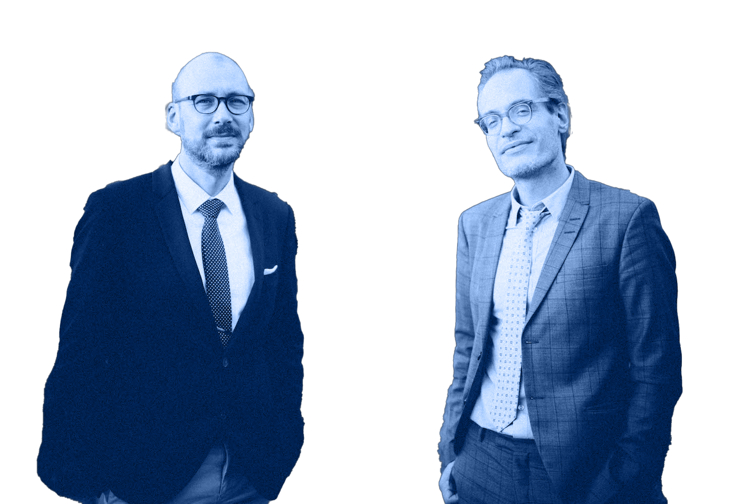
Pascal Baltzer and Matthias Dietzel
Outline
Concept
Objectives
Target audience
What our participants say
„
There is always something more to learn. Even for a master”, Master Oogway, Kung fu panda. This is absolutely the case with Pascal and Matthias at School Of Radiology #SOR, there’s always a place and new updated information for every radiologist from beginners to advanced levels. It’s always a pleasure attending thier webinars and symposiums, it’s not just a lecture as any lecture, they do it with absolute undeniable passion, pouring thier hearts out and most importantly they are always reachable just a message, an email or a chat in a coffee break away to answer all the questions and discuss even more. Best part for me would be the diveristy of cases, interactive long discussions, answering all the questions and inquires, sharing thier outstanding experiences in a friendly fun way, it’s never a dull moment with these two. As Rumi said “Stop learning. Start knowing”… start the journey of knowing with School Of Radiology.
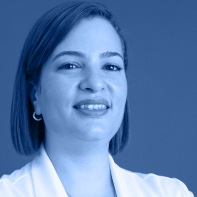
Dr. Menatallah Tawfik, M.D PhD
Dedicated consultant Breast imaging and Breast intervention radiologist. HOD Radiology & Breast unit at FHPC. National Breast Cancer Screening Program of UAE/EHS.
Programme
Time CET
Convert to your local time
09:00
Welcome
09:15
Why CEM?
- What information does CEM provide?
- In which clinical scenario does CEM make sense?
- What are the problems of CEM?
Practical implementation
09:30
Preparation and image acquisition
- What to consider before and during CEM?
- How to integrate CEM into your breast unit?
- Tips and tricks in practical implementation
10:15
Case discussion 1
- Artifacts
- Typical findings
- Typical limitations
10:45
Coffee break 1
11:00
Structured reporting
- Learn semantic BI-RADS criteria.
- Assess contrast enhancement.
- Are there decision rules?
11:45
Case discussion 2
- Apply structured reporting according to BI-RADS
- Distinguish typical benign from malignant lesions
- Understand the potential timing of image acquisition
13:00
Lunch break
13:45
Case discussion 3
- Integrate low energy and high energy image information
- See variations of malignant breast disease
- Recognize variations of benign breast disease
- Understand background parenchymal enhancement
Clinical application
15:00
Specific clinical scenarios
- Learn the potential value of CEM in specific clinical scenarios
- Understand the underlying evidence for CEM
- Management of CEM findings
15:45
Coffee break 2
16:00
Case discussion 4
- Value of CEM in specific clinical indications
- Typical findings in specific clinical indications
- Multimodal case interpretation in the clinical context
17:30
Wrap-up
17:45
End of the event
Literature
K. Coffey, M.S. Jochelson, Contrast-enhanced mammography in breast cancer screening, European Journal of Radiology. 156 (2022) 110513. https://doi.org/10.1016/j.ejrad.2022.110513
C.H. Lee, J. Phillips, J.S. Sung, J.M. Lewin, M.S. Newell, CONTRAST ENHANCED MAMMOGRAPHY (CEM) (A supplement to ACR BI-RADS® Mammography 2013), (2022). https://www.acr.org/-/media/ACR/Files/RADS/BI-RADS/BIRADS_CEM_2022.pdf
M.S. Jochelson, M.B.I. Lobbes, Contrast-enhanced Mammography: State of the Art, Radiology. 299 (2021) 36–48.
https://doi.org/10.1148/radiol.2021201948
Costs
Ticket
Price
Standard ticket
399,00 €
Flex Ticket
Your Course, Your Schedule!
With our Flex Tickets, you can secure your spot now without having to commit to a specific date right away. Perfect for those purchasing multiple tickets and looking to benefit from attractive bundle discounts.
How it works:
Purchase your ticket now, take advantage of bundle discounts, and let us know later which specific session you’d like to attend latter. This course is offered regularly. A simple email to us is all it takes to choose your date – flexible up to one hour before the session starts!
399,00 €
Discounts
Resident
-30%
Technician, radiographer
-75%
Bundle discount
-20%
You will automatically receive your bundle discount after placing an order containing more than one paid content.
Bundle discount may be combined with discounts “Resident”.
Venue: Online (Zoom)
Access links and technical instructions will be emailed to participants 3 days prior to the start of the event.
CME Accreditation
In our one day courses you will achieve optimal learning success and maximum CME points in the shortest possible time!
10
You want more?
Sure! If you book several courses at the same time, you receive a 20% discount on the total amount.
20
Resident in training?
We would like to make our contribution! We support your training with a 30% discount on all courses!
30
Quality assurance
This course is accredited by the Academy for Continuing Education of the German Radiological Society.
FAQ
What emails will I receive and when ?
You will receive all necessary documents from us by email.
The following information will be e-mailed to you:
- Immediately after your booking: booking confirmation + invoice + payment information (if you have chosen payment by invoice).
- 3 days before your course: Detailed information + access key to your course.
- Immediately after your course: Confirmation of participation.
Tip:
Please check the e-mail address that you provided for typos, and regularly check your inbox and junk mail folders.
I did not receive any e-mail from you!
Our emails are occasionally blocked by firewalls and junk mail filters. Unfortunately, we have no control over this. If you did not receive our e-mails, please reach out to us via e-mail, phone, instant messenger, or SMS:
We will gladly assist you and resend the information!
Tip:
Check your junk mail folder.
Add our address to your e-mail whitelist.
Check the e-mail you provided for typos.
How do I book a course?
All course bookings are done directly on our webshop.
Select your course(s) at https://school-of-radiology.com/courses/
Put the course(s) in your shopping basket.
Follow the instructions provided.
Tip:
If you have any problems, please feel free to contact us directly (see: “How can I contact you directly?”).
Do you have discounts?
Good news: You can get a wide range of discounts!
All discounts are applied automatically.
- Bundle discount:
You can receive a 20% bundle discount when booking a minimum of two courses at the same time. - Resident’s discount:
As a resident in training, you receive a 30% discount. - Combination:
All discounts can be combined.
Tip:
Many of our courses are sequential (basic + advanced courses).
You can book them together to receive your discount!
Even experts will benefit from joining our basic courses.
I have a voucher - how do I redeem it?
Here we briefly explain how you can redeem it.
How can I pay?
You can pay using a credit card, Paypal, or SEPA bank transfer. The amount will be debited automatically. We also accept manual payment by invoice.
Tip:
Our bank account information is not routinely included in the invoice.
Please copy the bank account details displayed at the end of the checkout process.
When will I receive my access code?
We will e-mail your access code to you 3 days before your course.
When will I receive my participation confirmation?
We will e-mail your participation confirmation immediately after your course.
In this file you will find:
- Your personal participation confirmation
- The number and confirmation of the CME credits you earned
- Confirmation of the number and types of cases you assessed during the course (this is optional; see also: “Will I receive a certificate on my case assessments?”)
Will I receive a certificate for my case assessments?
In our full-day courses, you will independently assess real-life cases. You will use our online tool and apply structured reporting for this assessment. Each case is quality-controlled, and we will discuss your assessment.
In our full-day courses we are happy to certify the number of cases that you independently reported (note: this service is optional).
In this case you will find the certificate in your participation confirmation on the last page.
Where can I find your bank details?
We display our bank account at the end of the checkout process. Note that our bank details are not included in the invoice.
Tip:
If you have any questions, please contact us directly (FAQ “How can I contact you directly?”).
How do I receive my CME credits?
We will e-mail your CME certificate directly after the course (see: “When will I receive my confirmation of participation?”). All the necessary information about the type and amount of your CME credits will be included.
Tip:
Details on the CME credits are outlined on each course site.
What technical equipment do you recommend?
Our courses are held online via Zoom.
All you need is an internet-compatible device of your choice and a stable internet connection.
The following tips can maximize your learning experience:
- Install Zoom on your computer.
- Log in with your personal Zoom account.
- Familiarize yourself with basic functionalities, e.g., turning video and audio on/off and writing chat messages.
- Please do not use Zoom via your browser.
- Tip: Install the Zoom app on a second device (smartphone, tablet, etc.). This will make answering interactive polls more convenient. It will also serve as a backup.
- Choose a high-quality monitor. If possible, use your diagnostic workstation.
- A fast and stable internet connection (ideally LAN, not W-LAN), appropriate lighting, a quality microphone, and a quality webcam are important elements for success in the course.
How can I contact you directly?
Our company does not have a hotline. We answer your questions personally.
You may also talk directly to your tutors for specific questions related to the course content.
So please contact us at any time by e-mail, phone, instant messenger, SMS, etc.:


